What Vitamins Should Not Be Taken With Rosuvastatin
A magnesium- and aluminum-containing antacid was reported to interfere with atorvastatin absorption. People can avoid this interaction by taking atorvastatin two hours before or after any aluminum/magnesium-containing antacids. Some magnesium supplements such as magnesium hydroxide are also antacids.
Membrane Fluidity In Alzheimer Disease
The role of the physicalchemical properties of intracellular membranous structures such as membrane fluidity in AD pathogenesis has been extensively studied. Membrane fluidity is a complex parameter, influenced both through some biophysical and biochemical factors . It is a parameter that reflects the main membrane characteristic organization . Experiments provide consistent data about membrane fluidity relations to various cellular processes, especially membrane processes. Changes in the membrane composition and structure could alter the conformation and function of transmembranal ion channels, as well as affect the interaction of receptors and effectors, leading to altered signal transduction, handling of Ca, and response to exogenous stimuli .
As shown in figure 1, we found a significant decrease of membrane fluidity in hippocampal neurons from AD patients compared with membranes from elderly non demented controls . Lower membrane uidity in AD patients was correlated with abnormal APP processing and cognitive decline .
Figure 6.
Filipin Staining And Image Analysis
Cells were fixed in PBS containing 4% paraformaldehyde for 30min, washed, and incubated with filipin for 2h at room temperature. Fluorescence imaging was performed on an Olympus IX-81 inverted microscope using a Nikon Intensilight illuminator, a Nikon Plan Apo VC 20× objective and a fluorescence filter set . Images were acquired with a CoolSnap K4 CCD camera via Micro-manager with the same acquisition settings across each experimental group. For analysis, three independent batches of cultures were analyzed . The total number of neurites or cells analyzed are reported . For the analysis of neurons, we manually selected ROIs covering neurites of morphologically identified neurons. Neurite selections were drawn to be approximately the same length for each ROI. For analysis of 3T3 cells, we used a threshold-based approach in ImageJ with a common threshold setting for all images in all experimental groups to select ROIs corresponding to cells. Average ROI intensity was measured in ImageJ. For every field of view , at least three ROIs from cell-free regions were manually selected, and their mean fluorescence intensities were calculated in the same manner. For background subtraction, the mean intensity value of every cell-containing ROI was subtracted by the average intensity of the three background ROIs in the same image.
Also Check: How Much Cholesterol In Canned Tuna
Preparations Of Human Pmnls
Procedures used for collecting blood samples from asymptomatic human volunteers were approved by the Institutional Review Board at either the University of Kentucky or the University of California, San Diego .
For some experiments, fresh blood was harvested by subcubital vein finger prick as described previously . After erythrocyte sedimentation at room temperature and 1 g for 45 min, the leukocyte-enriched plasma fraction containing neutrophils, monocytes, platelets, and sporadic erythrocytes was diluted 1:20 with Plasma-Lyte buffer containing 2.5 mM CaCl2 . To mimic the in vivo interactions between different types of blood cells, particularly for analyzing the shear-responses of nonadherent PMNLs, whole blood drawn into vacutainers using standard venipuncture was diluted 1:20 with Hanks’ buffered saline solution .
Changes In Cholesterol Levels In The Cell Membrane

To increase the levels of cholesterol in the neuron and in the plasma membrane, cells were incubated with media containing MCD/cholesterol complex . Unless otherwise stated, the concentration of cholesterol used in the experiments was 200 M. The cells were immediately incubated in this solution for 20 min at 37°C in culture medium and washed with PBS before adding A. To decrease the content of cholesterol, cells were incubated with MCD . Unless otherwise stated, the concentration used in the experiments was 3 mM. Cells were incubated for 30 min at 37°C with this solution in culture medium. Subsequently, the cells were washed with PBS and A treatments were initiated. It is important to consider that both MCD and MCD/Cholesterol complexes likely modify the cholesterol content in internal membranes. Therefore, to quantify the changes in the membrane, we used Fillipin III that is believed to interact mainly with membrane sterols . In addition, with ANEP GP imaging Staining and GP Imaging), we only considered pixels in the peripheral plasma membrane during the analysis .
Don’t Miss: Is Shrimp Bad For Your Cholesterol
Heterogeneity In Membrane Physical Property
Discrete lipid domains with differing composition, and thus membrane fluidity, can coexist in model lipid membranes this can be observed using fluorescence microscopy. The biological analogue, ‘lipid raft‘, is hypothesized to exist in cell membranes and perform biological functions. Also, a narrow annular lipid shell of membrane lipids in contact with integral membrane proteins have low fluidity compared to bulk lipids in biological membranes, as these lipid molecules stay stuck to surface of the protein macromolecules.
How Does Cholesterol Affect The Fluidity Of A Plasma Membrane
I was previously taught that cholesterol affects the fluidity of a plasma membrane. At high temperatures, cholesterol decreases fluidity and at low temperatures cholesterol increases fluidity. The Khan academy and Wikipedia pages below say the same thing.
Yet, in a current college course, the textbook says that cholesterol reduces the mobility of the first few CH2 groups of a phospholipid’s two fatty acid chains. In this way, the cholesterol makes the lipid bilayer more rigid and decreases the lipid bilayer’s permeability to small, water-soluble molecules. However, it says that cholestrol does not actually make the membrane less fluid.
Is the textbook from my current course just a more nuanced explanation, or have I misunderstood something else?
Also Check: Does Pasta Have High Cholesterol
How Does Cholesterol Affect The Fluidity And Permeability Of Cell Membranes
Cholesterol interacts with the fatty acid tails of phospholipids to moderate the properties of the membrane: Cholesterol functions to immobilise the outer surface of the membrane, reducing fluidity. It makes the membrane less permeable to very small water-soluble molecules that would otherwise freely cross.
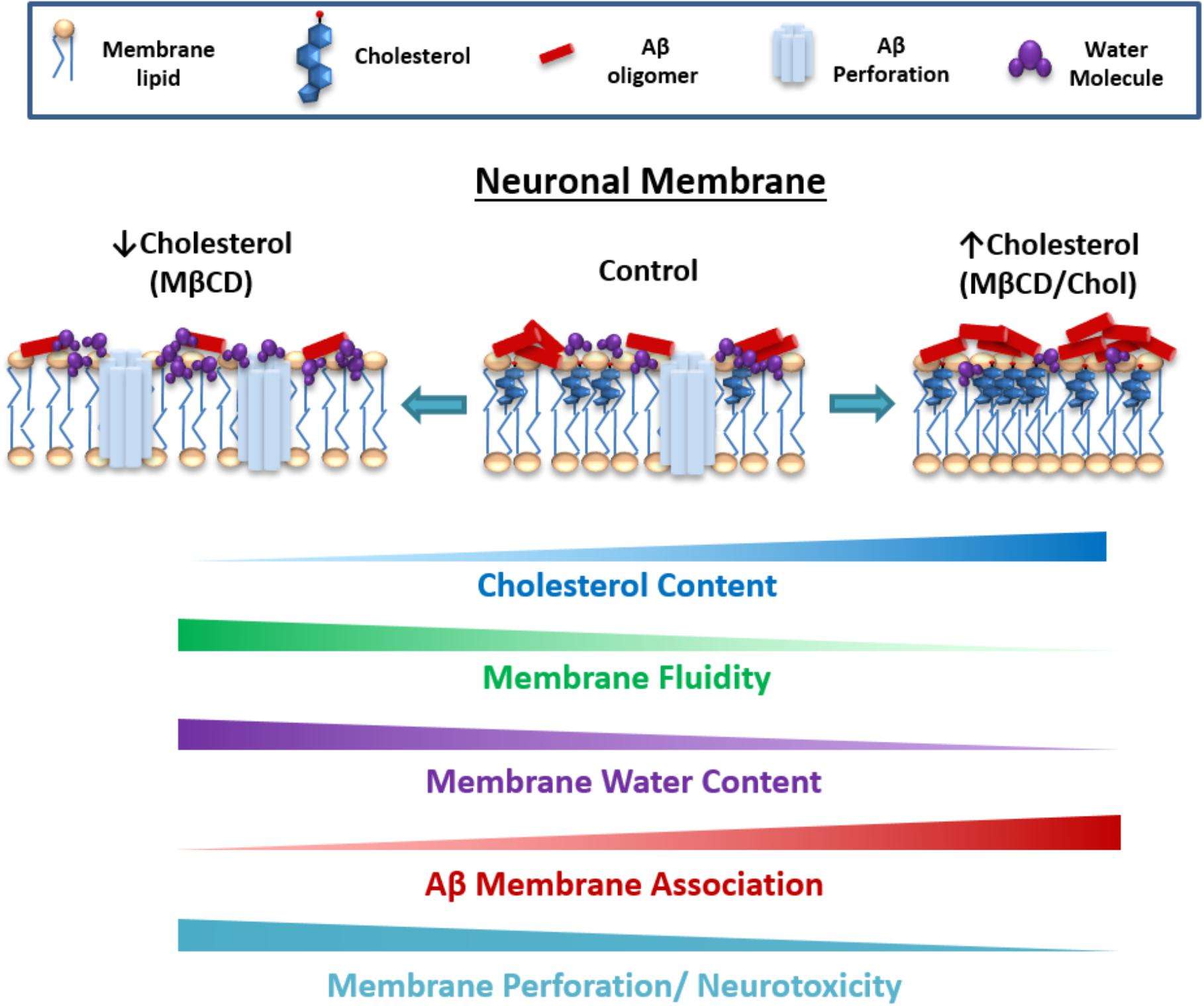
Increase In Cholesterol Reduced The Membrane Perforation Induced By A
FIGURE 4. The effect of A on membrane integrity depends on the level of cholesterol. Representative traces of capacitive currents recorded at different times after increasing or decreasing relative levels of cholesterol in HEK-293 cells. Time course of A-induced perforation for each of the conditions described in showing the kinetic of the perforation process and how this effect changes when membrane-cholesterol levels are modified. Quantification of membrane charge transferred at the end of the registration period. The effects of A in a cholesterol-depleted condition were blocked by the NA7 peptide. Bars represent the mean ± SEM. Asterisk denotes p< 0.005.
You May Like: Is Canned Tuna Good For Lowering Cholesterol
Role Of Dietary Lipids In Alzheimer Disease
Recent theories suggests that there would be an interaction between genetic predisposition and environmental factors that lead to cell death by amyloid toxicity or disruption of tau protein. Dietary lipids could be a determining factor in the difference in risk between developed and underdeveloped countries. Dietary lipids could be the primary risk factor in late-onset sporadic AD . The critical factors seem to be the ratios of polyunsaturated fatty acids to monounsaturated , saturated fatty acids to essential fatty acids . These contents are modified by the APOE4 genotype .
Lipid lowering agents appear to have a protective effect, although studies are not conclusive. Statins decrease the oxidizability of LDL, with decreased levels of oxygen reactive species, anti-inflammatory effects and improve endothelial dysfunction, also increased alpha-secretase activity. Increase the synthesis of LDL receptors, with decreased circulating level and reduced production of PPA.
The histological changes seen in the initial stages of AD confirmed that membrane lipids and inflammation are involved in the disease . AGE n-3/n-6 rate has a major impact on the balance of eicosanoid metabolism inflammatory and anti-inflammatory, and the degree of saturation of membrane lipids and fluidity affects its function. The apoE4 genotype may influence the risk of AD, as it is unable to protect that transports lipids from oxidation .
Figure 7.
Myelin And Cell Membrane Fluidity
Cell membrane fluidity is a parameter describing the freedom of movement of protein and lipid constituents within the cell membrane. CMF appears to influence several cellular processes including the activity of membrane-associated enzymes.68,69 CMF may also be implicated in the changes associated with the aging process. Age-associated lowering of D6D activity will decrease PGE1 synthesis,70 which would be expected to increase CMF.71 The activity of membrane-associated enzymes increases in fluid membranes.68 Among the significant components of cell membranes are the phospholipids that contain FAs. Phospholipids made from SFAs have a different structure and are less fluid than those that incorporate an EFA. LA and ALA have an effect on the neuronal CMF. They are able to decrease the cholesterol level in the neuronal membrane, which would decrease membrane fluidity, which in turn would make it difficult for the cell to carry out its normal functions and increase the cells susceptibility to injury and death.67 Higher membrane USFA levels are associated with increased CMF.72
Andras Balajthy, … Zoltan Varga, in, 2017
Read Also: Is Oyster High In Cholesterol
Factor #: Cholesterol Content Of The Bilayer
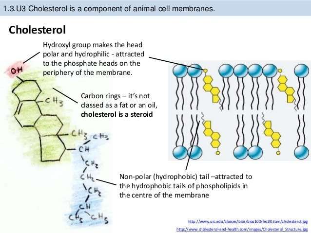
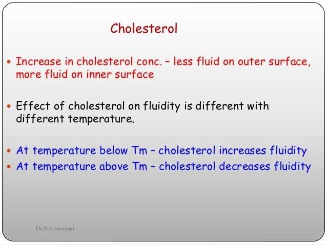
Cholesterol has a somewhat more complicated relationship with membrane fluidity. You can think of it is a buffer that helps keep membrane fluidity from getting too high or too low at high and low temperatures.
At low temperatures, phospholipids tend to cluster together, but steroids in the phospholipid bilayer fill in between the phospholipids, disrupting their intermolecular interactions and increasing fluidity.
At high temperatures, the phospholipids are further apart. In this case, cholesterol in the membrane has the opposite effect and pulls phospholipids together, increasing intermolecular forces and decreasing fluidity.
Cell Lysis And Immunoblottings

Cells were lysed with the RIPA buffer TritonX-100, 0.1% SDS, 1% sodium deoxycholate) supplemented with mammalian protease and phosphatase inhibitor cocktails as described previously . Protein concentrations were determined via BCA protein assays . In general, 2030g of protein samples were loaded per lane onto SDS-PAGE gels to be resolved, before transferred to nitrocellulose membranes . Membranes were then blocked with 4% non-fat-milk in PBS at room temperature for an hour, followed by incubation with primary antibodies at 4°C overnight. The blots were washed with PBS for three times before incubation with secondary antibodies. Membranes were finally washed with PBS and detected on a LICOR Odyssey system. Captured images were quantitated using Image Studio software as per manufacturers instructions.
Read Also: Does Honey Nut Cheerios Really Lower Cholesterol
Cholesterol Recognition Sites May Mediate The Sterolprotein Interactions Of Kv13
Fig. 4. Schematic representation of the cholesterol recognition motifs identified along the Kv1.3 sequence. Each subunit contains at least seven putative cholesterol recognition sites. Two CARC sequences are located on the distal end of the C-terminus. As we previously demonstrated removal of the last 84 amino acid residues abolishes cholesterol sensitivity of Kv1.3.
Fig. 5. The effect of 420 M CHOL loading on the gating properties of various Kv1.3 constructs. CHO cells were transiently transfected with the following constructs: wild-type Kv1.3, CKv1.3 , a CARC4 mutant , and a CARC5 mutant and loaded with 420 M CHOL. To determine the activation time constant and the midpoint of the steady-state activation we applied the same protocols as we discussed in Fig. 7. This figure demonstrates the relative change in the activation time constant and V1/2 due to the cholesterol loading in case of a particular construct.
Wanda M. Haschek, … Matthew A. Wallig, in, 2010
Pmnl Responses To Shear Stress Depend On The Cholesterol
The attenuating effects of membrane cholesterol enrichment on PMNL shear-responses depended on the concentration of cholesterol:MCD used to preincubate PMNLs . Increasing the concentration of cholesterol complexes reduced PMNL responses to shear dose-dependently. When incubated with 10 g/ml cholesterol:MCD, the shear-responses of PMNLs were reversed. Concomitantly, incubation with increasing cholesterol:MCD decreased the fluidity of plasma membranes in a similar dose-dependent fashion . Particularly, when cholesterol complexes were greater than 1 g/ml, membrane fluidity was significantly lower than that of naïve cells.
Fig. 4.Dose-dependent impairment of PMNL shear-responses by membrane-modifying cholesterol:MCD conjugates correlates with their effects on cell membrane fluidity. A: PMNL shear-responses were determined for cells pretreated with 010 g/ml cholesterol:MCD and 10 nM FMLP followed by exposure to 5 dyn/cm2 shear stress for 10 min. n = 4 . *P< 0.002 compared with samples without CH treatment. B: membrane fluidity for cells incubated with 010 g/ml cholesterol:MCD was measured and normalized to that of untreated cells. n 3. #P< 0.001 compared with samples without CH treatment. C: linear regression analysis for PMNL shear-responses and membrane fluidity. PRR indices for cells pretreated with 1 and 2 g/ml CH were included from prior experiments .
Read Also: Beer Increases Cholesterol
How Cholesterol Affects The Health Of The Body
Cholesterol circulates in your blood as the level of cholesterol increases, so does the risk of your health. This is why you need to keep a check on your cholesterol levels as frequently as possible.
There are two types of cholesterol: LDL cholesterol, which is terrible for your health, and HDL, which is essential for your health. Therefore, you should know that too much LDL cholesterol or too low HDL levels will increase the risk for your body.
This will slowly lead to the build-up of cholesterol in the arteries inner walls that feed the brain and heart. Cholesterol can also join the other substance to form a thick deposit on the side of the arteries.
This will narrow the arteries and make them less flexible this condition is termed as atherosclerosis.
If, for instance, a blood clot is formed, blocking one of these narrowed arteries could cause a stroke or and heart attack. High cholesterol levels are one of the crucial controllable risk factors for coronary heart disease, Heart attack, and stroke.
The chance of risk can increase if you smoke, have high blood or diabetes. The viscosity of the lipid bilayer of a cell membrane or a synthetic lipid membrane is known as membrane fluidity.
The packing of lipids influences the fluidity of the membrane. The membrane fluidity is also affected by fatty acids the unsaturated or saturated nature of fatty acids affects the membrane fluids.
The Inevitable Question Is Quo Vadis
The salient signalling pathways which S100A4 activates encompass growth factor receptors in the regulation of cell proliferation and the cell division cycle and the apoptotic pathway, among others. On the metastasis front, the participation of S100A4 in cancer invasion by ECM remodelling, alteration of cell membrane fluidity, and in the modulation cell cytoskeletal functional dynamics is well established, so is its link with neovascularisation. The discussion of these attributes underscores the viability of appropriate strategy for therapeutic approach to target it. S100A4 is a normal gene, not exclusively linked with cancer but deregulated in its expression leading to the expression of the cancer phenotype.
The quest for targets for therapeutic manipulation has to fulfil several criteria. Is the postulated target a biomarker for tumour invasion, growth and metastatic progression? Is the maker widely expressed in human cancers? Do the signalling pathways of the potential target lend themselves to pharmacological manipulation? The answer to these queries is arguably yes. If so, much pre-clinical work is needed with potential inhibitors as single agents or in combination with conventional cytotoxic agents. This could lay the groundwork to pose these questions in clinical trials.
Christopher Hillis, Wendy Lim, in, 2018
Don’t Miss: Is Tuna High In Cholesterol
Increased Membrane Cholesterol Levels Augmented Association Of A Aggregates In Hippocampal Neurons
To obtain a better understanding of cholesterol influence on the A membrane association, we performed the opposite experiment . The neurons treated with cholesterol showed a significant increase in A association compared with the control condition . Interestingly, we also found some large aggregates associated with the neuronal tissue . The analysis of the segmented data showed that the cluster number and size increased in the presence of supplemental cholesterol . For instance, in cholesterol supplemented neurons, we found an increase of 45% in the number of A clusters compared to those obtained in control conditions : 44.1 ± 3.6 Figure 3E). Interestingly, the difference was particularly more significant when we quantified the area of these clusters . For example, the data revealed that cluster area in control conditions was 0.18 ± 0.01 vs. an average value of 0.46 ± 0.05 in MCD-cholesterol treatment , representing an approximately 150% increase in cluster area due to the treatment with cholesterol as compared to control .
Why Do Unsaturated Fats Increase Membrane Fluidity
If unsaturated fatty acids are compressed, the kinks in their tails push adjacent phospholipid molecules away, which helps maintain fluidity in the membrane. Cholesterol functions as a buffer, preventing lower temperatures from inhibiting fluidity and preventing higher temperatures from increasing fluidity.
You May Like: Does Shrimp Have Good Cholesterol
How Does Lipid Composition Affect Membrane Fluidity
Lipid composition has no effect on the fluidity of membranes. Unsaturated fatty acids tend to make the membrane less fluid because kinks introduced by the double bonds keep them from packing together well. Sterols, such as cholesterol, can either increase or decrease membrane fluidity depending on temperature.
Immunocytochemistry And Image Analysis

For immunostaining after electrophysiological recordings, coverslips containing recorded cells were fixed in PBS containing 4% paraformaldehyde, washed, blocked for 1h with PBS containing 1% BSA, and incubated overnight at 4°C with diluted primary antibodies . Secondary antibodies with distinct fluorophores were then incubated at room temperature for 2h. Fluorescence imaging was performed on an Olympus IX-51 inverted microscope with a 60× UPlanFL objective and a Flash 4.0 sCMOS camera . The optical filter sets for Alexa 488, 568, and 647 fluorescence were, respectively: Ex 470/20 DiC 510LP 535/25, Ex 565/25 DiC 585LP Em 630/90, and Ex 630/60 DiC 660LP Em 695/100. For each fluorescence channel, images were taken with the same acquisition settings . Biocytin-positive neurons were positioned approximately in the center of the fields of view for imaging. Not all electrophysiologically recorded neurons were identified and imaged, as some were damaged during Biocytin infusion.
Also Check: How Much Cholesterol In Crab