Cholesterol Accessibility At The Ciliary Membrane Controls Hedgehog Signaling
- Open annotations. The current annotation count on this page is being calculated.
Defective Lamellipodial Formation In Cholestenone Containing Cells
We noted that the coase-treated cells extended long filopodia-like protrusions toward the wounded area . To elucidate some of the early changes accompanying the non-migratory phenotype of coase-treated cells, we analysed actin in HDFs at 3 h post wounding. Phalloidin staining revealed that in coase treated cells the actin rich protrusions at the leading edge were very narrow and spindle shaped, whereas control and MBCD-treated cells exhibited prominent well spread lamellae . At this time point, the average width of the lamellipodium in coase-treated cells was reduced by over 50% . Lamellipodium spreading depends on the actin branch point complex Arp2/3 . We found that the Arp2/3 complex component ARPC2 was efficiently recruited to the lamellipodia of control and MBCD-treated cells at 3 h post treatment . In contrast, the coase-treated cells displayed a prominent cytosolic ARPC2 staining, and reduced immunostaining at the leading edge . These results suggest that actin dynamics is aberrantly regulated in the presence of cholestenone. This agrees with earlier reports showing that coase treatment affects the diffusion of membrane lipids and proteins in an actin dependent manner , .
Purification And Labeling Of Lipid Probes
Mutant His6-tagged Perfringolysin O was purified as previously described and covalently labeled with Alexa Fluor 647 following the manufacturer’s instructions. Expression, purification and labeling of His6-tagged OlyA and His6-tagged OlyA_E69A was carried out as described previously . Briefly, both proteins were expressed in Escherichia coli RosettapLysS cells, purified by metal-affinity and gel-filtration chromatography and their lone cysteines labelled with Atto-647 maleimide following the manufacturerâs instructions . Expression and purification of His6- and FLAG-tagged domain 4 of anthrolysin O was carried out as described previously .
Don’t Miss: Does Feta Cheese Affect Cholesterol
Cell Culture And Drug Treatments
NIH/3T3-CG Reporter Cells used in the CRISPR screen , Smo-/- MEFs stably expressing SMO mutants , NIH/3T3 Flp-In cells stably expressing GPR161-YFP, mouse HM1 mESCs, and Ptch1-/- cells have been previously described and characterized . NIH/3T3 Flp-In cells were purchased from Thermo Fisher Scientific, NIH/3T3 cells from ATCC, and HM1 mESCs from Open Biosystems and used at low passages for experiments. These purchased cells lines came with a certificate of authentication from the vendor and were used without further validation. Cell lines were confirmed to be negative for Mycoplasma infection. In the NIH/3T3 Flp-In background, Lss-/-, Dhcr7-/- and Dhcr24-/- clonal cell lines were generated using a two-cut CRISPR strategy using methods described in our previous publications and validated using a PCR-based genotyping strategy . NIH/3T3 cells and Ptch-/- MEFs stably expressing ARL13B C-terminally tagged with GFP were generated by lentiviral infection followed by puromycin selection.
Background: Brain Biology 101

Although I have tried to write this essay in a way that is accessibleto the non-expert, it will still be helpful to first familiarize youwith basic knowledge of the structure of the brain and the rolesplayed by different cell types within the brain.
In addition to the neurons, the brain also contains a large number of”helper” cells called glial cells, which are concerned with the careand feeding of neurons. Three principle types of glial cells willplay a role in our later discussion: the microglia, the astrocytes,and the oligodendrocytes. Microglia are the equivalent of white bloodcells in the rest of the body. They are concerned with fighting offinfective agents such as bacteria and viruses, and they also monitorneuron health, making life-and-death decisions: programming a particularneuron for apoptosis if it appears to bemalfunctioning beyond hope of recovery, or is infected with anorganism that is too dangerous to let flourish.
Also Check: Is Deer Meat High In Cholesterol
Accessible Cholesterol Regulates Hedgehog Signaling
As an orthogonal approach to test the importance of accessible cholesterol without using myriocin, we used the cholesterol binding domain of the bacterial toxin anthrolysin O . Unlike a reagent like MβCD, which extracts cholesterol from cells, ALOD4 selectively traps accessible cholesterol on the outer leaflet of the plasma membrane without altering cholesterol levels in the cell or plasma membrane . Despite very different mechanisms of action, both ALOD4 and MβCD blocked HH signaling when added to cells . In a control experiment, ALOD4 induced the expression of Hmgcr, the gene encoding the enzyme HMG-CoA reductase, which is known to be induced when accessible cholesterol is depleted . ALOD4 did not change the frequency of ciliation .
ALOD4 impairs hedgehog signaling by trapping accessible cholesterol.
In summary, HH signaling is enhanced by myriocin, which increases accessible cholesterol , but inhibited by ALOD4, which decreases accessible cholesterol. Neither myriocin nor ALOD4 change total cholesterol abundance in cells . We conclude that accessible cholesterol is the thermodynamically distinct fraction of total cholesterol that is relevant for the regulation of SMO in HH signaling.
Enzymes In The Cholesterol Biosynthesis Pathway Positively Regulate Hedgehog Signaling
Enzymes that generate cholesterol positively regulate hedgehog signaling.
The post-squalene portion of the cholesterol biosynthetic pathway, with enzymes colored according to their FDR corrected p-value in our CRISPR screens . Two branches of the pathway produce cholesterol, while a third shunt pathway produces 24, 25-epoxycholesterol. DHCR24 is the only enzyme that is required for cholesterol biosynthesis, but dispensable for 24, 25-epoxycholesterol synthesis. HH signaling strength in Lss-/-, Dhcr7-/- and Dhcr24-/- NIH/3T3 cells was assessed by measuring Gli1 mRNA by quantitative reverse transcription PCR after treatment with either HiSHH or HiSHH combined with 0.3 mM cholesterol:MβCD complexes. Bars denote the mean value derived from the four individual measurements shown. Statistical significance was determined by the Mann-Whitney test or the Kruskal-Wallis test .
In summary, the data from our genetic screen support the view that cholesterol itself, rather than a precursor or a metabolite, is the endogenous sterol lipid that regulates SMO activation. Caveats of genetic screens include their inability to identify genes or pathways that are redundant, required for cell viability or growth, or dependent on non-enzymatic reactions or exogenous molecules supplied by the media.
Read Also: Is Shrimp Bad For Your Cholesterol
Excessive Oxygen Exposure And Cognitive Decline
It has been observed that some elderly people suffer temporary andsometimes permanent cognitive decline following a lengthy operation.Researchers at the University of South Florida and VanderbiltUniversity suspected that this might be due to excessive exposure tooxygen . Typically, during an operation, people are often administeredhigh doses of oxygen, even as much as 100% oxygen. The researchersconducted an experiment on young adult mice, which had been engineeredto be predisposed towards Alzheimer’s but had not yet sufferedcognitive decline. They did however already have amyloid-betadeposits in their brains. The re-engineered mice, as well as acontrol group that did not have the Alzheimer’s susceptibility gene,were exposed to 100-percent oxygen for a period of three hours, threetimes over the course of several months, simulating repeated operations. They found that the Alzheimer’s pre-disposed mice suffered significantcognitive decline following the oxygen exposure, by contrast with thecontrol mice.
How Can I Raise My Hdl Level
If your HDL level is too low, lifestyle changes may help. These changes may also help prevent other diseases, and make you feel better overall:
- Eat a healthy diet. To raise your HDL level, you need to eat good fats instead of bad fats. This means limiting saturated fats, which include full-fat milk and cheese, high-fat meats like sausage and bacon, and foods made with butter, lard, and shortening. You should also avoid trans fats, which may be in some margarines, fried foods, and processed foods like baked goods. Instead, eat unsaturated fats, which are found in avocado, vegetable oils like olive oil, and nuts. Limit carbohydrates, especially sugar. Also try to eat more foods naturally high in fiber, such as oatmeal and beans.
- Stay at a healthy weight. You can boost your HDL level by losing weight, especially if you have lots of fat around your waist.
- Exercise. Getting regular exercise can raise your HDL level, as well as lower your LDL. You should try to do 30 minutes of moderate to vigorous aerobic exercise on most, if not all, days.
- Avoid cigarettes.Smoking and exposure to secondhand smoke can lower your HDL level. If you are a smoker, ask your health care provider for help in finding the best way for you to quit. You should also try to avoid secondhand smoke.
- Limit alcohol. Moderate alcohol may lower your HDL level, although more studies are needed to confirm that. What we do know is that too much alcohol can make you gain weight, and that lowers your HDL level.
You May Like: Is Shrimp Bad For Your Cholesterol
Is The Fluid Mosaic A Suitable Model To Describe Fundamental Features Of Biological Membranes What May Be Missing
- 1Center for Biomembrane Physics , University of Southern Denmark, Odense, Denmark
- 2Membrane Biophysics and Biophotonics group, Department of Biochemistry and Molecular Biology, University of Southern Denmark, Odense, Denmark
- 3Department of Physics, Chemistry, and Pharmacy, University of Southern Denmark, Odense, Denmark
The structure, dynamics, and stability of lipid bilayers are controlled by thermodynamic forces, leading to overall tensionless membranes with a distinct lateral organization and a conspicuous lateral pressure profile. Bilayers are also subject to built-in curvature-stress instabilities that may be released locally or globally in terms of morphological changes leading to the formation of non-lamellar and curved structures. A key controller of the bilayers propensity to form curved structures is the average molecular shape of the different lipid molecules. Via the curvature stress, molecular shape mediates a coupling to membrane-protein function and provides a set of physical mechanisms for formation of lipid domains and laterally differentiated regions in the plane of the membrane. Unfortunately, these relevant physical features of membranes are often ignored in the most popular models for biological membranes. Results from a number of experimental and theoretical studies emphasize the significance of these fundamental physical properties and call for a refinement of the fluid mosaic model .
In Summary: Structure Of The Cell Membrane
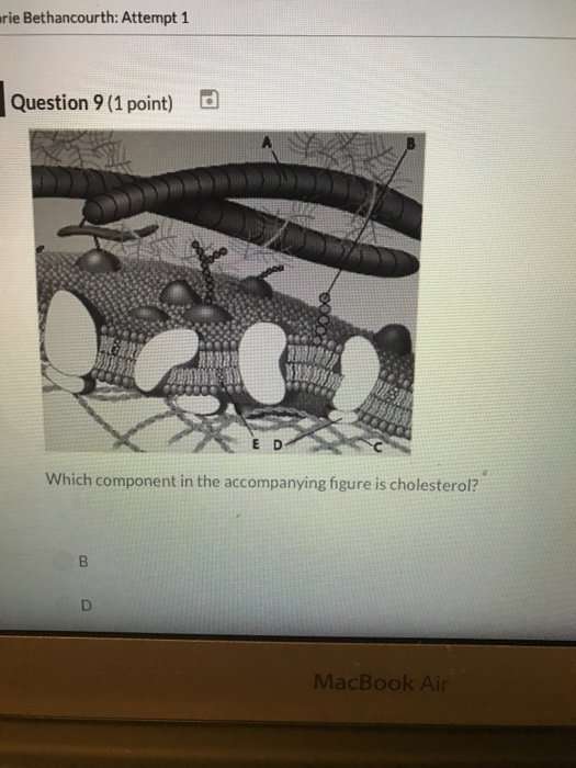
The modern understanding of the plasma membrane is referred to as the fluid mosaic model. The plasma membrane is composed of a bilayer of phospholipids, with their hydrophobic, fatty acid tails in contact with each other. The landscape of the membrane is studded with proteins, some of which span the membrane. Some of these proteins serve to transport materials into or out of the cell. Carbohydrates are attached to some of the proteins and lipids on the outward-facing surface of the membrane. These form complexes that function to identify the cell to other cells. The fluid nature of the membrane owes itself to the configuration of the fatty acid tails, the presence of cholesterol embedded in the membrane , and the mosaic nature of the proteins and protein-carbohydrate complexes, which are not firmly fixed in place. Plasma membranes enclose the borders of cells, but rather than being a static bag, they are dynamic and constantly in flux.
Recommended Reading: Does Pork Have High Cholesterol
Establishing A Laboratory Testing Process
Important activities for establishing a routine method of analysis are shown in the accompanying figure. The blocks at the bottom illustrate the key steps involved in routine analysis, where the laboratory acquires specimens, performs tests, checks statistical QC, and reports test results. Those activities are generally regarded as the real work of the laboratory.
However, for analysis to become routine, the other activities shown in the figure are very important. The selection of the diagnostic test is actually the first step, but this is often skipped for common tests whose medical usefulness is well accepted. For these well accepted tests, we usually start with the selection of the method , then validate its performance. If performance is acceptable, the method is implemented for routine service. If performance is not acceptable, the laboratory may develop some improvements, although that is becoming increasingly difficult with the high state of automation of many analytical systems. Today it’s more likely that a laboratory would select another method rather than attempt to make improvements, then start the validation process over again for the new method.
We invite you to read the rest of this article.
Fatty Acids And Triacylglycerides

The fatty acids are lipids that contain long-chain hydrocarbons terminated with a carboxylic acid functional group. Because the long hydrocarbon chain, fatty acids are hydrophobic or nonpolar. Fatty acids with hydrocarbon chains that contain only single bonds are called saturated fatty acids because they have the greatest number of hydrogen atoms possible and are, therefore, saturated with hydrogen. Fatty acids with hydrocarbon chains containing at least one double bond are called unsaturated fatty acids because they have fewer hydrogen atoms. Saturated fatty acids have a straight, flexible carbon backbone, whereas unsaturated fatty acids have kinks in their carbon skeleton because each double bond causes a rigid bend of the carbon skeleton. These differences in saturated versus unsaturated fatty acid structure result in different properties for the corresponding lipids in which the fatty acids are incorporated. For example, lipids containing saturated fatty acids are solids at room temperature, whereas lipids containing unsaturated fatty acids are liquids.
A triacylglycerol, or triglyceride, is formed when three fatty acids are chemically linked to a glycerol molecule . Triglycerides are the primary components of adipose tissue , and are major constituents of sebum . They play an important metabolic role, serving as efficient energy-storage molecules that can provide more than double the caloric content of both carbohydrates and proteins.
Recommended Reading: Can Keto Cause High Cholesterol
What Gets Stored In A Cookie
This site stores nothing other than an automatically generated session ID in the cookie no other information is captured.
In general, only the information that you provide, or the choices you make while visiting a web site, can be stored in a cookie. For example, the site cannot determine your email name unless you choose to type it. Allowing a website to create a cookie does not give that or any other site access to the rest of your computer, and only the site that created the cookie can read it.
A Focused Crispr Screen Targeting Lipid
CRISPR screens identify lipid-related genes that influence hedgehog signaling.
Flowchart summarizing the screening strategy. Screens for positive and negative regulators used a high, saturating concentration of SHH or a low concentration of SHH , respectively. Volcano plots of the HiSHH-Bot10% screen for positive regulators and the LoSHH-Top5% screen for negative regulators. Enrichment is calculated as the mean of all sgRNAs for a given gene in the sorted over unsorted population, with the y-axis showing significance based on the false discovery rate -corrected p-value. Screen results analyzed by grouping genes based on the core lipid biosynthetic pathways in KEGG. In all panels, genes identified as positive and negative regulators are labeled in blue and orange respectively. See Supplementary file 4 for the full analysis.
The screens correctly identified all four positive controls included in the library: Smo and Adrbk1 as positive regulators and Ptch1 and Sufu as negative regulators . In addition, genes previously known to influence HH signaling and protein trafficking at primary cilia were amongst the most significant hits . In addition to Inpp5e, other genes involved in phosphoinositide metabolism were also significant hits. Pla2g3, which encodes a secreted phospholipase, was identified as a negative regulator of HH signaling, an effect that may be related to its known role as a suppressor of ciliogenesis .
Read Also: Are Mussels High In Cholesterol
Cellular Sphingomyelin Suppresses Hedgehog Signaling
Multiple enzymes in the sphingolipid synthesis pathway were statistically significant hits in the LoSHH-Top5% screen, indicating these enzymes are negative regulators of HH signaling strength . Top hits from this screen include Sptlc2, the first committed step in sphingolipid synthesis from L-serine and palmitoyl-CoA, as well as Sgms1, which converts ceramide to sphingomyelin . The identification of Sgms1 suggests that SM is the relevant product of the sphingolipid pathway that attenuates HH signaling.
Enzymes that generate sphingomyelin negatively regulate hedgehog signaling.
Several control experiments established that the potentiating effect of myriocin on HH signaling was caused by the depletion of SM, rather than an unrelated effect. First, fumonisin B1, a mycotoxin structurally distinct from myriocin that inhibits a different step in SM synthesis , also amplified HH signaling . Second, the potentiating effect of myriocin on HH signaling could be reversed by the exogenous administration of SM . Finally, increasing SM levels in cells using a low-dose of staurosporine had the expected opposite effect: reduction of HH signaling strength .
New Challenges And Future Perspectives
Are there examples from naturally occurring membranes displaying micrometer-sized domains as observed in model membrane systems? Yes, in very specialized membranes such as lung surfactant and skin stratum corneum, where lipids are the principal components, membrane-cytoskeleton anchorage is lacking, and local equilibrium conditions are likely attainable , cf. Figure 2. Other examples have been reported, such as platelets upon activation , macrophages , T-cells , yeast , red blood cells , fibroblasts although it is not yet clear whether these observations are controlled by the same mechanisms. The message here is that generalizations can be perilous, and it is probably a good idea to pay attention to the compositional diversity of different membranes, including the way that processes evolve .
Read Also: Do Cheerios Really Lower Cholesterol